Information on COVID-19, Kidney Disease, and Telemedicine.

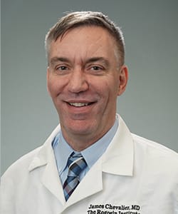
James Chevalier, M.D.

Specialties:

  • Nephrology

Expertise:

  • Nephrotic Syndrome
  • Glomerulonephritis, including lupus nephritis, minimal change disease, membranous nephropathy, IgA nephropathy
  • Polycystic Kidney Disease

Board Certifications:

  • Nephrology
  • Internal Medicine

Clinical and Academic Appointments:

  • Director, Jack J. Dreyfus Kidney Clinic
  • Assistant Professor of Medicine and Medicine in Surgery, Weill Cornell Medicine
  • Assistant Attending Physician, NewYork-Presbyterian Hospital

Education and Training:

  • Medical School:  University at Buffalo School of Medicine and Biomedical Sciences
  • Residency:  NewYork-Presbyterian/Weill Cornell
  • Fellowship in Nephrology: NewYork-Presbyterian/Weill Cornell

Locations:

Rogosin Manhattan East Dialysis
505 East 70th Street
New York, NY 10021
212-746-1566
Get Directions+

Publications

  • Automatically Measuring Kidney, Liver, and Cyst Volumes in Autosomal Dominant Polycystic Kidney Disease
    Qing Xiong, Xinzi He, Elisa Scalco, Siria Pasini, Chenglin Zhu, Mina C Moghadam, Usama Sattar, Vahid Davoudi, Vahid Bazojoo, Hreedi Dev, Mengjun Shen, Zhongxiu Hu, Sophie Shih, Serena J Prince, Jon D Blumenfeld, Robert J Min, James M Chevalier, Daniil Shimonov, Rebecca J Lepping, Alan S L Yu, Mert R Sabuncu, Anna Caroli, Martin R Prince...

    J Am Soc Nephrol. 2025 Nov 4. doi: 10.1681/ASN.0000000904. Online ahead of print.

    ABSTRACT

    BACKGROUND: Kidney, liver and cyst volumes are important for diagnosis, classification and management of autosomal dominant polycystic kidney disease (ADPKD) but challenging to measure accurately and reproducibly. Here, we develop a web-based deep learning platform to automatically and robustly measure kidneys, liver and cyst volumes in ADPKD.

    METHODS: MRI and CT scans from ADPKD patients (n=611) and participants without ADPKD (n=109) were used to train a 3D hybrid model combining U-Net and transformer elements for segmenting kidneys, liver and cysts. The model is implemented as a web-based calculator at www.traceorg.com, providing segmentation labels, volumes and Mayo Clinic Image Classification (MIC). Automatic browser anonymization of DICOM images ensures privacy. Internal validation was conducted on 70 MRIs for kidney and liver segmentations, 46 MRIs for cyst segmentations and performance was compared to 5 open access segmentation models (TotalSegmentator, MR Annotator, Kim, Woznicki and Gregory-Kline). External validation was performed on one single-center dataset (n=58), one multicenter dataset (n=73), CRISP2 (n=30) and PKD-RRC (n=115) MRIs with T2-weighted and T1-weighted images.

    RESULTS: After training on 720 participants (mean age=48±15, eGFR=74±32 ml/min/1.73m2 and htTKV=826±772ml/m), TraceOrg internal validation performance achieved high mean Dice scores of 0.97 (kidneys), 0.97 (liver), 0.93 (kidney cysts) and 0.82 (liver cysts) outperforming existing models for ADPKD. External validation showed strong performance with Dice scores of 0.92-0.94 (kidney), 0.87-0.96 (liver), 0.85 (kidney cysts) and 0.76-0.90 (liver cysts) for the single-center and 0.95 (kidney), 0.81 (kidney cysts) for the multicenter dataset. Compared to CRISP volumes measured by stereology, mean absolute percent difference was 5.3% (kidneys, n=30), 11% (kidney cysts, n=30) and 5.5% (liver, n=22). Compared to PKD-RRC (n=115), mean absolute percent difference in TKV was 4.9%.

    CONCLUSIONS: TraceOrg is a publicly available web-based tool that automatically measures kidney, liver and cyst volumes from abdominal MRI in ADPKD with high accuracy compared to manual segmentations.

    PMID:41186985 | DOI:10.1681/ASN.0000000904

  • Deep learning-based liver cyst segmentation in MRI for autosomal dominant polycystic kidney disease
    Mina Chookhachizadeh Moghadam, Mohit Aspal, Xinzi He, Dominick J Romano, Arman Sharbatdaran, Zhongxiu Hu, Kurt Teichman, Hui Yi Ng He, Usama Sattar, Chenglin Zhu, Hreedi Dev, Daniil Shimonov, James M Chevalier, Akshay Goel, George Shih, Jon D Blumenfeld, Mert R Sabuncu, Martin R Prince...

    Radiol Adv. 2024 May 23;1(2):umae014. doi: 10.1093/radadv/umae014. eCollection 2024 Jul.

    ABSTRACT

    BACKGROUND: Autosomal dominant polycystic kidney disease (ADPKD) can lead to polycystic liver disease (PLD), characterized by liver cysts. Although majority of the patients are asymptomatic, massively enlarged liver secondary to PLD can cause discomfort, and compression on adjacent structures requiring cyst aspiration/fenestration, partial liver resection, or liver transplantation. Monitoring PLD by measuring liver volume fails to track the early stages when liver cyst volume is too small to affect liver volume.

    PURPOSE: To improve PLD assessment in the early stages by automating detection and segmentation of liver cysts using deep learning (DL) models.

    MATERIALS AND METHODS: A self-configured UNet-based platform (nnU-Net) was trained with 40 ADPKD subjects with liver cysts annotated by a radiologist. Internal (n = 7), External (n = 10), and test-retest reproducibility (n = 17) validations included macro- and micro-level performance metrics: patient-level Dice scores (PDice), along with voxel-level true positive rates (VTPR), as well as analysis of time saved in a model-assisted scenario. Additionally, we assessed human-level reliability in liver cyst segmentation and evaluated the model's test-retest reproducibility. We further compared liver volume vs cyst volume for tracking disease in a subject with 16+ years follow-up.

    RESULTS: The model achieved an 82% ± 11% PDice and a 75% ± 15% VTPR on the internal test sets (n = 7 patients), and 80% ± 12% Dice score and a 91% ± 7% VTPR on the external test sets (n = 10 patients). It excelled particularly in detecting small liver cysts, a challenging task for manual annotation. This efficiency translated to a median of 91% (IQR: 14%) reduction in annotation time compared to manual labeling. Test-retest assessment demonstrated excellent reproducibility, with coefficients of variation of 94% for liver cyst fraction and 92% for cyst count.

    CONCLUSION: DL automation of liver cyst segmentations demonstrates potential to improve tracking of liver cyst volume in polycystic liver disease.

    PMID:41059391 | PMC:PMC12429238 | DOI:10.1093/radadv/umae014

  • Reporting ADPK Disease Phenotypes on Abdominal Scans
    Zhongxiu Hu, Elizabeth G Lane, Grace C Lo, Jon D Blumenfeld, Daniil Shimonov, James M Chevalier, Emily A Schonfeld, Danielle Brandman, Martin R Prince...

    Kidney Int Rep. 2025 Jul 1;10(9):2967-2976. doi: 10.1016/j.ekir.2025.06.046. eCollection 2025 Sep.

    ABSTRACT

    INTRODUCTION: Kidney and liver volumes from abdominal magnetic resonance imaging (MRI) and computed tomography (CT) scans are critical biomarkers recommended by Kidney Disease: Improving Global Outcomes (KDIGO) for autosomal dominant polycystic kidney disease (ADPKD) progression and response to therapy. The purpose of this study was to determine how often these biomarkers are included in radiology reports as well as their reproducibility.

    METHODS: Outside abdominal MRI (n = 102) and CT (n = 43) studies were reviewed retrospectively and independently by 2 observers for prevalence of reporting ADPKD-relevant findings with discrepancies resolved by 2 radiologists. These 2 radiologists independently reevaluated all examinations to assess interobserver reproducibility.

    RESULTS: Outside reports (n = 145; males: 46%; median age: 47 [interquartile range: 35-61] years) by 122 radiologists from 88 institutions included kidney volumes in only 30 (21%) reports. Out of 140 imaging examinations that included the entire liver, 1 (1%) outside report provided liver volume. Comparison of outside and study radiologists' kidney volume measurements showed a median absolute difference of 15%, using the ellipsoidal method and 8% using model-assisted contouring. Additional positive findings not mentioned in 145 outside reports included umbilical hernia (n = 44), hepatic steatosis (n = 3), inguinal hernia (n = 4), pancreatic cyst (n = 6) and severe inferior vena cava (IVC) compression by cysts (n = 2).

    CONCLUSION: Radiologists report reliably on complex cysts, calcifications, ascites, and abdominal aortic aneurysms in ADPKD. However, the clinical utility of these reports can be improved more reliably by including kidney volume and other important imaging features of ADPKD recommended by clinical guidelines.

    PMID:40980645 | PMC:PMC12446943 | DOI:10.1016/j.ekir.2025.06.046

  • Effects of Pregnancy on Liver and Kidney Cyst Growth Rates in Autosomal Dominant Polycystic Kidney Disease: A Pilot Study
    Vahid Bazojoo, Vahid Davoudi, Jon D Blumenfeld, Chenglin Zhu, Line Malha, Grace C Lo, James M Chevalier, Daniil Shimonov, Arman Sharbatdaran, Hreedi Dev, Syed I Raza, Zhongxiu Hu, Xinzi He, Arindam RoyChoudhury, Martin R Prince...

    J Clin Med. 2025 May 24;14(11):3688. doi: 10.3390/jcm14113688.

    ABSTRACT

    Background/Objectives: Polycystic liver disease (PLD) is the most common extrarenal manifestation of autosomal dominant polycystic kidney disease (ADPKD). PLD is more prevalent in women, and women have larger liver cysts, possibly due to estrogen-related mechanisms. Maternal estrogen levels normally increase during pregnancy. Thus, we investigated the pregnancy-associated increase in liver volume, liver cyst volume, total kidney volume (TKV), and kidney cyst growth rates in ADPKD patients. Methods: Kidney, liver, and cyst volumes were measured in 16 ADPKD patients by magnetic resonance imaging (MRI) at multiple timepoints before and after pregnancy. The log-transformed TKV, liver volume, and cyst volume growth rates during a period with pregnancy were compared to a period without pregnancy. Results: In ADPKD patients, a higher annualized liver cyst growth rate was observed during a period with pregnancy compared to a period without pregnancy (34 ± 16%/yr vs. 23 ± 17%/yr; p-value = 0.005). Liver volume growth was also higher during a period with pregnancy, 6 [2, 7]%/yr vs. 0.3 [-0.4, 2]%/yr (p-value = 0.04). In addition, the mean kidney cyst growth rate was higher (12 ± 11%/yr vs. 4 ± 9%/yr; p-value = 0.05), and there was a trend toward a pregnancy-associated increase in the TKV growth rate (6 [4, 8]%/yr vs. 3 [0.8, 5]%/yr, (p-value = 0.14) during a period with pregnancy. Conclusions: In patients with ADPKD, the liver volume and cyst volume growth rates increased during pregnancy. This supports the hypothesis that the estrogen-mediated stimulation of liver cyst growth may contribute to the severe polycystic liver disease that is more prevalent in women than men with ADPKD. Further studies with larger populations are needed to explore the mechanisms and long-term implications of these findings.

    PMID:40507450 | PMC:PMC12156408 | DOI:10.3390/jcm14113688

  • The Role of Baseline Total Kidney Volume Growth Rate in Predicting Tolvaptan Efficacy for ADPKD Patients: A Feasibility Study
    Hreedi Dev, Zhongxiu Hu, Jon D Blumenfeld, Arman Sharbatdaran, Yelynn Kim, Chenglin Zhu, Daniil Shimonov, James M Chevalier, Stephanie Donahue, Alan Wu, Arindam RoyChoudhury, Xinzi He, Martin R Prince...

    J Clin Med. 2025 Feb 21;14(5):1449. doi: 10.3390/jcm14051449.

    ABSTRACT

    Background/Objectives: Although tolvaptan efficacy in ADPKD has been demonstrated in randomized clinical trials, there is no definitive method for assessing its efficacy in the individual patient in the clinical setting. In this exploratory feasibility study, we report a method to quantify the change in total kidney volume (TKV) growth rate to retrospectively evaluate tolvaptan efficacy for individual patients. Treatment-related changes in estimated glomerular filtration rate (eGFR) are also assessed. Methods: MRI scans covering at least 1 year prior to and during treatment with tolvaptan were performed, with deep learning facilitated kidney segmentation and fitting multiple imaging timepoints to exponential growth in 32 ADPKD patients. Clustering analysis differentiated tolvaptan treatment "responders" and "non-responders" based upon the magnitude of change in TKV growth rate. Differences in rate of eGFR decline, urine osmolality, and other parameters were compared between responders and non-responders. Results: Eighteen (56%) tolvaptan responders (mean age 42 ± 8 years) were identified by k-means clustering, with an absolute reduction in annual TKV growth rate of >2% (mean = -5.1% ± 2.5% per year). Thirteen (44%) non-responders were identified, with <1% absolute reduction in annual TKV growth rate (mean = +2.4% ± 2.7% per year) during tolvaptan treatment. Compared to non-responders, tolvaptan responders had significantly higher mean TKV growth rates prior to tolvaptan treatment (7.1% ± 3.6% per year vs. 3.7% ± 2.4% per year; p = 0.003) and higher median pretreatment spot urine osmolality (Uosm, 393 mOsm/kg vs. 194 mOsm/kg, p = 0.03), confirmed by multivariate analysis. Mean annual rate of eGFR decline was less in responders than in non-responders (-0.25 ± 0.04, CI: [-0.27, -0.23] mL/min/1.73 m2 per year vs. -0.40 ± 0.06, CI: [-0.43, -0.37] mL/min/1.73 m2 per year, p = 0.036). Conclusions: In this feasibility study designed to assess predictors of tolvaptan treatment efficacy in individual patients with ADPKD, we found that high pretreatment levels of annual TKV growth rate and higher pretreatment spot urine osmolality were associated with a responder phenotype.

    PMID:40094908 | PMC:PMC11899928 | DOI:10.3390/jcm14051449

  • Improved predictions of total kidney volume growth rate in ADPKD using two-parameter least squares fitting
    Zhongxiu Hu, Arman Sharbatdaran, Xinzi He, Chenglin Zhu, Jon D Blumenfeld, Hanna Rennert, Zhengmao Zhang, Andrew Ramnauth, Daniil Shimonov, James M Chevalier, Martin R Prince...

    Sci Rep. 2024 Jun 14;14(1):13794. doi: 10.1038/s41598-024-62776-8.

    ABSTRACT

    Mayo Imaging Classification (MIC) for predicting future kidney growth in autosomal dominant polycystic kidney disease (ADPKD) patients is calculated from a single MRI/CT scan assuming exponential kidney volume growth and height-adjusted total kidney volume at birth to be 150 mL/m. However, when multiple scans are available, how this information should be combined to improve prediction accuracy is unclear. Herein, we studied ADPKD subjects ( n = 36 ) with 8+ years imaging follow-up (mean = 11 years) to establish ground truth kidney growth trajectory. MIC annual kidney growth rate predictions were compared to ground truth as well as 1- and 2-parameter least squares fitting. The annualized mean absolute error in MIC for predicting total kidney volume growth rate was 2.1 % ± 2 % compared to 1.1 % ± 1 % ( p = 0.002 ) for a 2-parameter fit to the same exponential growth curve used for MIC when 4 measurements were available or 1.4 % ± 1 % ( p = 0.01 ) with 3 measurements averaging together with MIC. On univariate analysis, male sex ( p = 0.05 ) and PKD2 mutation ( p = 0.04 ) were associated with poorer MIC performance. In ADPKD patients with 3 or more CT/MRI scans, 2-parameter least squares fitting predicted kidney volume growth rate better than MIC, especially in males and with PKD2 mutations where MIC was less accurate.

    PMID:38877066 | PMC:PMC11178802 | DOI:10.1038/s41598-024-62776-8

  • A Primer for Utilizing Deep Learning and Abdominal MRI Imaging Features to Monitor Autosomal Dominant Polycystic Kidney Disease Progression
    Chenglin Zhu, Xinzi He, Jon D Blumenfeld, Zhongxiu Hu, Hreedi Dev, Usama Sattar, Vahid Bazojoo, Arman Sharbatdaran, Mohit Aspal, Dominick Romano, Kurt Teichman, Hui Yi Ng He, Yin Wang, Andrea Soto Figueroa, Erin Weiss, Anna G Prince, James M Chevalier, Daniil Shimonov, Mina C Moghadam, Mert Sabuncu, Martin R Prince...

    Biomedicines. 2024 May 20;12(5):1133. doi: 10.3390/biomedicines12051133.

    ABSTRACT

    Abdominal imaging of autosomal dominant polycystic kidney disease (ADPKD) has historically focused on detecting complications such as cyst rupture, cyst infection, obstructing renal calculi, and pyelonephritis; discriminating complex cysts from renal cell carcinoma; and identifying sources of abdominal pain. Many imaging features of ADPKD are incompletely evaluated or not deemed to be clinically significant, and because of this, treatment options are limited. However, total kidney volume (TKV) measurement has become important for assessing the risk of disease progression (i.e., Mayo Imaging Classification) and predicting tolvaptan treatment's efficacy. Deep learning for segmenting the kidneys has improved these measurements' speed, accuracy, and reproducibility. Deep learning models can also segment other organs and tissues, extracting additional biomarkers to characterize the extent to which extrarenal manifestations complicate ADPKD. In this concept paper, we demonstrate how deep learning may be applied to measure the TKV and how it can be extended to measure additional features of this disease.

    PMID:38791095 | PMC:PMC11118119 | DOI:10.3390/biomedicines12051133

  • Test Retest Reproducibility of Organ Volume Measurements in ADPKD Using 3D Multimodality Deep Learning
    Xinzi He, Zhongxiu Hu, Hreedi Dev, Dominick J Romano, Arman Sharbatdaran, Syed I Raza, Sophie J Wang, Kurt Teichman, George Shih, James M Chevalier, Daniil Shimonov, Jon D Blumenfeld, Akshay Goel, Mert R Sabuncu, Martin R Prince...

    Acad Radiol. 2024 Mar;31(3):889-899. doi: 10.1016/j.acra.2023.09.009. Epub 2023 Oct 3.

    ABSTRACT

    RATIONALE AND OBJECTIVES: Following autosomal dominant polycystic kidney disease (ADPKD) progression by measuring organ volumes requires low measurement variability. The objective of this study is to reduce organ volume measurement variability on MRI of ADPKD patients by utilizing all pulse sequences to obtain multiple measurements which allows outlier analysis to find errors and averaging to reduce variability.

    MATERIALS AND METHODS: In order to make measurements on multiple pulse sequences practical, a 3D multi-modality multi-class segmentation model based on nnU-net was trained/validated using T1, T2, SSFP, DWI and CT from 413 subjects. Reproducibility was assessed with test-re-test methodology on ADPKD subjects (n = 19) scanned twice within a 3-week interval correcting outliers and averaging the measurements across all sequences. Absolute percent differences in organ volumes were compared to paired students t-test.

    RESULTS: Dice similarlity coefficient > 97%, Jaccard Index > 0.94, mean surface distance < 1 mm and mean Hausdorff Distance < 2 cm for all three organs and all five sequences were found on internal (n = 25), external (n = 37) and test-re-test reproducibility assessment (38 scans in 19 subjects). When averaging volumes measured from five MRI sequences, the model automatically segmented kidneys with test-re-test reproducibility (percent absolute difference between exam 1 and exam 2) of 1.3% which was better than all five expert observers. It reliably stratified ADPKD into Mayo Imaging Classification (area under the curve=100%) compared to radiologist.

    CONCLUSION: 3D deep learning measures organ volumes on five MRI sequences leveraging the power of outlier analysis and averaging to achieve 1.3% total kidney test-re-test reproducibility.

    PMID:37798206 | PMC:PMC10957335 | DOI:10.1016/j.acra.2023.09.009

  • Clinical Quality Control of MRI Total Kidney Volume Measurements in Autosomal Dominant Polycystic Kidney Disease
    Chenglin Zhu, Hreedi Dev, Arman Sharbatdaran, Xinzi He, Daniil Shimonov, James M Chevalier, Jon D Blumenfeld, Yi Wang, Kurt Teichman, George Shih, Akshay Goel, Martin R Prince...

    Tomography. 2023 Jul 12;9(4):1341-1355. doi: 10.3390/tomography9040107.

    ABSTRACT

    Total kidney volume measured on MRI is an important biomarker for assessing the progression of autosomal dominant polycystic kidney disease and response to treatment. However, we have noticed that there can be substantial differences in the kidney volume measurements obtained from the various pulse sequences commonly included in an MRI exam. Here we examine kidney volume measurement variability among five commonly acquired MRI pulse sequences in abdominal MRI exams in 105 patients with ADPKD. Right and left kidney volumes were independently measured by three expert observers using model-assisted segmentation for axial T2, coronal T2, axial single-shot fast spin echo (SSFP), coronal SSFP, and axial 3D T1 images obtained on a single MRI from ADPKD patients. Outlier measurements were analyzed for data acquisition errors. Most of the outlier values (88%) were due to breathing during scanning causing slice misregistration with gaps or duplication of imaging slices (n = 35), slice misregistration from using multiple breath holds during acquisition (n = 25), composing of two overlapping acquisitions (n = 17), or kidneys not entirely within the field of view (n = 4). After excluding outlier measurements, the coefficient of variation among the five measurements decreased from 4.6% pre to 3.2%. Compared to the average of all sequences without errors, TKV measured on axial and coronal T2 weighted imaging were 1.2% and 1.8% greater, axial SSFP was 0.4% greater, coronal SSFP was 1.7% lower and axial T1 was 1.5% lower than the mean, indicating intrinsic measurement biases related to the different MRI contrast mechanisms. In conclusion, MRI data acquisition errors are common but can be identified using outlier analysis and excluded to improve organ volume measurement consistency. Bias toward larger volume measurements on T2 sequences and smaller volumes on axial T1 sequences can also be mitigated by averaging data from all error-free sequences acquired.

    PMID:37489475 | PMC:PMC10366880 | DOI:10.3390/tomography9040107

  • Effect of Averaging Measurements From Multiple MRI Pulse Sequences on Kidney Volume Reproducibility in Autosomal Dominant Polycystic Kidney Disease
    Hreedi Dev, Chenglin Zhu, Arman Sharbatdaran, Syed I Raza, Sophie J Wang, Dominick J Romano, Akshay Goel, Kurt Teichman, Mina C Moghadam, George Shih, Jon D Blumenfeld, Daniil Shimonov, James M Chevalier, Martin R Prince...

    J Magn Reson Imaging. 2023 Oct;58(4):1153-1160. doi: 10.1002/jmri.28593. Epub 2023 Jan 16.

    ABSTRACT

    BACKGROUND: Total kidney volume (TKV) is an important biomarker for assessing kidney function, especially for autosomal dominant polycystic kidney disease (ADPKD). However, TKV measurements from a single MRI pulse sequence have limited reproducibility, ± ~5%, similar to ADPKD annual kidney growth rates.

    PURPOSE: To improve TKV measurement reproducibility on MRI by extending artificial intelligence algorithms to automatically segment kidneys on T1-weighted, T2-weighted, and steady state free precession (SSFP) sequences in axial and coronal planes and averaging measurements.

    STUDY TYPE: Retrospective training, prospective testing.

    SUBJECTS: Three hundred ninety-seven patients (356 with ADPKD, 41 without), 75% for training and 25% for validation, 40 ADPKD patients for testing and 17 ADPKD patients for assessing reproducibility.

    FIELD STRENGTH/SEQUENCE: T2-weighted single-shot fast spin echo (T2), SSFP, and T1-weighted 3D spoiled gradient echo (T1) at 1.5 and 3T.

    ASSESSMENT: 2D U-net segmentation algorithm was trained on images from all sequences. Five observers independently measured each kidney volume manually on axial T2 and using model-assisted segmentations on all sequences and image plane orientations for two MRI exams in two sessions separated by 1-3 weeks to assess reproducibility. Manual and model-assisted segmentation times were recorded.

    STATISTICAL TESTS: Bland-Altman, Schapiro-Wilk (normality assessment), Pearson's chi-squared (categorical variables); Dice similarity coefficient, interclass correlation coefficient, and concordance correlation coefficient for analyzing TKV reproducibility. P-value < 0.05 was considered statistically significant.

    RESULTS: In 17 ADPKD subjects, model-assisted segmentations of axial T2 images were significantly faster than manual segmentations (2:49 minute vs. 11:34 minute), with no significant absolute percent difference in TKV (5.9% vs. 5.3%, P = 0.88) between scans 1 and 2. Absolute percent differences between the two scans for model-assisted segmentations on other sequences were 5.5% (axial T1), 4.5% (axial SSFP), 4.1% (coronal SSFP), and 3.2% (coronal T2). Averaging measurements from all five model-assisted segmentations significantly reduced absolute percent difference to 2.5%, further improving to 2.1% after excluding an outlier.

    DATA CONCLUSION: Measuring TKV on multiple MRI pulse sequences in coronal and axial planes is practical with deep learning model-assisted segmentations and can improve TKV measurement reproducibility more than 2-fold in ADPKD.

    EVIDENCE LEVEL: 2 TECHNICAL EFFICACY: Stage 1.

    PMID:36645114 | PMC:PMC10947493 | DOI:10.1002/jmri.28593

  • Deep Learning Automation of Kidney, Liver, and Spleen Segmentation for Organ Volume Measurements in Autosomal Dominant Polycystic Kidney Disease
    Arman Sharbatdaran, Dominick Romano, Kurt Teichman, Hreedi Dev, Syed I Raza, Akshay Goel, Mina C Moghadam, Jon D Blumenfeld, James M Chevalier, Daniil Shimonov, George Shih, Yi Wang, Martin R Prince...

    Tomography. 2022 Jul 13;8(4):1804-1819. doi: 10.3390/tomography8040152.

    ABSTRACT

    Organ volume measurements are a key metric for managing ADPKD (the most common inherited renal disease). However, measuring organ volumes is tedious and involves manually contouring organ outlines on multiple cross-sectional MRI or CT images. The automation of kidney contouring using deep learning has been proposed, as it has small errors compared to manual contouring. Here, a deployed open-source deep learning ADPKD kidney segmentation pipeline is extended to also measure liver and spleen volumes, which are also important. This 2D U-net deep learning approach was developed with radiologist labeled T2-weighted images from 215 ADPKD subjects (70% training = 151, 30% validation = 64). Additional ADPKD subjects were utilized for prospective (n = 30) and external (n = 30) validations for a total of 275 subjects. Image cropping previously optimized for kidneys was included in training but removed for the validation and inference to accommodate the liver which is closer to the image border. An effective algorithm was developed to adjudicate overlap voxels that are labeled as more than one organ. Left kidney, right kidney, liver and spleen labels had average errors of 3%, 7%, 3%, and 1%, respectively, on external validation and 5%, 6%, 5%, and 1% on prospective validation. Dice scores also showed that the deep learning model was close to the radiologist contouring, measuring 0.98, 0.96, 0.97 and 0.96 on external validation and 0.96, 0.96, 0.96 and 0.95 on prospective validation for left kidney, right kidney, liver and spleen, respectively. The time required for manual correction of deep learning segmentation errors was only 19:17 min compared to 33:04 min for manual segmentations, a 42% time saving (p = 0.004). Standard deviation of model assisted segmentations was reduced to 7, 5, 11, 5 mL for right kidney, left kidney, liver and spleen respectively from 14, 10, 55 and 14 mL for manual segmentations. Thus, deep learning reduces the radiologist time required to perform multiorgan segmentations in ADPKD and reduces measurement variability.

    PMID:35894017 | PMC:PMC9326744 | DOI:10.3390/tomography8040152

  • Bortezomib for Reduction of Proteinuria in IgA Nephropathy
    Choli Hartono, Miriam Chung, Alan S Perlman, James M Chevalier, David Serur, Surya V Seshan, Thangamani Muthukumar...

    Kidney Int Rep. 2018 Mar 11;3(4):861-866. doi: 10.1016/j.ekir.2018.03.001. eCollection 2018 Jul.

    ABSTRACT

    INTRODUCTION: IgA nephropathy is the most common glomerulonephritis in the world. We conducted a pilot trial (NCT01103778) to test the effect of bortezomib in patients with IgA nephropathy and significant proteinuria.

    METHODS: We treated 8 consecutive subjects from July 2011 until March 2016 with 4 doses of bortezomib. All subjects had biopsy-proven IgA nephropathy and proteinuria of greater than 1 g per day. They were given 4 doses of bortezomib i.v. at 1.3 mg/m2 of body surface area per dose. Changes in proteinuria and renal function were followed for 1 year after enrollment. The primary endpoint was full remission defined as proteinuria of less than 300 mg per day.

    RESULTS: All 8 subjects received and tolerated 4 doses of bortezomib over a 2-week period during enrollment. The median baseline daily proteinuria was 2.46 g (interquartile range: 2.29-3.16 g). At 1-year follow-up, 3 subjects (38%) had achieved the primary endpoint. The 3 subjects who had complete remission had Oxford classification T scores of 0 before enrollment. Of the remaining 5 subjects, 1 was lost to follow-up within 1 month of enrollment and 4 (50%) did not have any response or had progression of disease.

    CONCLUSION: Proteasome inhibition by bortezomib may reduce significant proteinuria in select cases of IgA nephropathy. Subjects who responded to bortezomib had Oxford classification T score of 0 and normal renal function.

    PMID:29988921 | PMC:PMC6035125 | DOI:10.1016/j.ekir.2018.03.001

  • Multiparametric Quantitative Ultrasound Imaging in Assessment of Chronic Kidney Disease
    Jing Gao, Alan Perlman, Safa Kalache, Nathaniel Berman, Surya Seshan, Steven Salvatore, Lindsey Smith, Natasha Wehrli, Levi Waldron, Hanish Kodali, James Chevalier...

    J Ultrasound Med. 2017 Nov;36(11):2245-2256. doi: 10.1002/jum.14209. Epub 2017 Apr 13.

    ABSTRACT

    OBJECTIVES: To evaluate the value of multiparametric quantitative ultrasound imaging in assessing chronic kidney disease (CKD) using kidney biopsy pathologic findings as reference standards.

    METHODS: We prospectively measured multiparametric quantitative ultrasound markers with grayscale, spectral Doppler, and acoustic radiation force impulse imaging in 25 patients with CKD before kidney biopsy and 10 healthy volunteers. Based on all pathologic (glomerulosclerosis, interstitial fibrosis/tubular atrophy, arteriosclerosis, and edema) scores, the patients with CKD were classified into mild (no grade 3 and <2 of grade 2) and moderate to severe (at least 2 of grade 2 or 1 of grade 3) CKD groups. Multiparametric quantitative ultrasound parameters included kidney length, cortical thickness, pixel intensity, parenchymal shear wave velocity, intrarenal artery peak systolic velocity (PSV), end-diastolic velocity (EDV), and resistive index. We tested the difference in quantitative ultrasound parameters among mild CKD, moderate to severe CKD, and healthy controls using analysis of variance, analyzed correlations of quantitative ultrasound parameters with pathologic scores and the estimated glomerular filtration rate (GFR) using Pearson correlation coefficients, and examined the diagnostic performance of quantitative ultrasound parameters in determining moderate CKD and an estimated GFR of less than 60 mL/min/1.73 m2 using receiver operating characteristic curve analysis.

    RESULTS: There were significant differences in cortical thickness, pixel intensity, PSV, and EDV among the 3 groups (all P < .01). Among quantitative ultrasound parameters, the top areas under the receiver operating characteristic curves for PSV and EDV were 0.88 and 0.97, respectively, for determining pathologic moderate to severe CKD, and 0.76 and 0.86 for estimated GFR of less than 60 mL/min/1.73 m2 . Moderate to good correlations were found for PSV, EDV, and pixel intensity with pathologic scores and estimated GFR.

    CONCLUSIONS: The PSV, EDV, and pixel intensity are valuable in determining moderate to severe CKD. The value of shear wave velocity in assessing CKD needs further investigation.

    PMID:28407281 | PMC:PMC5640470 | DOI:10.1002/jum.14209

  • Serum Inflammatory and Immune Mediators Are Elevated in Early Stage Diabetic Nephropathy
    Alan S Perlman, James M Chevalier, Patrick Wilkinson, Hao Liu, Thomas Parker, Daniel M Levine, Betty Jo Sloan, Anna Gong, Raymond Sherman, Francis X Farrell...

    Ann Clin Lab Sci. 2015 Spring;45(3):256-63.

    ABSTRACT

    BACKGROUND: Diabetes is the leading cause of end stage renal disease (ESRD) in the United States, representing 44% of incident cases [1]. In this study, serum and peripheral blood collected from diabetic patients in five stages of chronic kidney disease (CKD), as defined by glomerular filtration rate (GFR), were compared to healthy (non-CKD) subjects.

    METHODS: Serum samples were analyzed for 39 inflammatory or immune mediator protein levels and peripheral blood samples were analyzed for expression of 35 gene transcripts.

    RESULTS: In serum, MCP-1, FGF-2, VEGF, and EGF levels were elevated above controls at all stages of DN. Five mediator levels, GM-CSF, IL-1α, IL-1RA, IL-6, and MIP1β increased with disease progression until stage 4-5, at which point a decrease was observed paralleling a loss of functional renal mass that occurs in late stage CKD. Five mediator levels: GRO, IFNγ, MDC, Eotaxin, and G-CSF significantly differed from controls at one or more stages without apparent correlation with disease stage. Only a single mediator, sIL2RA, exhibited a linear increase with disease severity consistent with declining GFR. In peripheral blood, the transcript level of seven mediators, ICAM1, TNF-α, TGF-β, IL-8, IL17RA, IFNγ, and MYD88 were significantly elevated at all disease stages as compared to control.

    CONCLUSION: Statistically significant differences in protein and transcripts levels between diseased and control can be detected in serum and peripheral blood utilizing high content profiling. These changes occur as early as stage 1-2 before a significant decline in renal function.

    PMID:26116588

Request an Appointment

Specialties

Request an Appointment

For your convenience, a representative from The Rogosin Institute can contact you to schedule an appointment. Please complete the necessary information.